Scaled reference drawings of architeuthis?

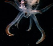
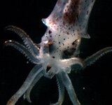
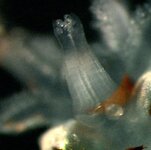
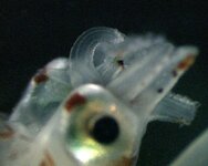
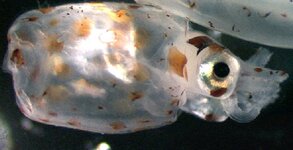
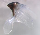
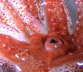
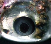
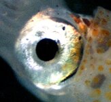
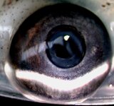
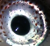
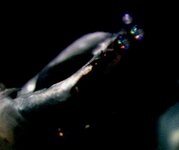
These are of juvenile Moroteuthis (ML ~ 15mm), taken beneath a microscope (very difficult to focus on a moving animal this small, beneath a scope).

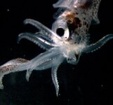
It'll give you some indication what the buccal membrane/beaks are doing, and how far they protrude from the arms of the animal.

Moroteuthis and Architeuthis are VERY similar beasts (if you forget about the adult hooks on the tentacles, and warty skin of Moroteuthis).
 

Attachments

  • conv_287523.jpg
    conv_287523.jpg
    69.4 KB · Views: 136
  • conv_287524.jpg
    conv_287524.jpg
    77.6 KB · Views: 138
  • conv_287525.jpg
    conv_287525.jpg
    78.2 KB · Views: 132
Here's a few of larval Nototodarus (the rhynchoteuthis stage ... a true larva, rather than paralarve for the majority of other squid and octopus); although you can't see the beaks/buccal membrane on tehse, they might be of greater interest to you Jean (am posting them here rather than starting a new thread).

I've long-since forgotten the camera specs, but there are so many systems on the market now that this sort of image could be obtained by the majority (you just want to limit the objective lens to 0.32-1x).

The entire larval Nototodarus (probably 5-6mm TL)


Close up of head of larval Nototodarus

Close up of fused tentacles (proboscis)
 

Attachments

  • conv_287528.jpg
    conv_287528.jpg
    84.8 KB · Views: 102
  • conv_287527.jpg
    conv_287527.jpg
    81.3 KB · Views: 154
  • conv_287526.jpg
    conv_287526.jpg
    53.9 KB · Views: 153
Terrific shots and very helpful, thanks Steve :smile:

The multiple views of the beak with the ruler are just what I needed. 8)

If I’m interpreting the images of Moroteuthis correctly, it looks like the lower beak is nearly transparent and the buccal membrane is extending beyond the beak tip on the ventral side?

--Carl
:ammonite:
 
CarlS said:
If I’m interpreting the images of Moroteuthis correctly, it looks like the lower beak is nearly transparent and the buccal membrane is extending beyond the beak tip on the ventral side?

It's subject to some interpretation Carl; indeed the beaks of paralarval forms are nearly transparent (the hood darkens first), and on the earlier images are probably surrounded by the buccal lips rather than buccal membrane; without the exact specimen before me I couldn't say for sure.

This is an even closer image of the beak, lip etc. in paralarval Moroteuthis (I didn't know I had this shot myself ... been dredging through the old files ...); the view is from the lower (ventral) surface; that's the funnel to the left of the image; the ventral arms are quite poorly developed ona paralarva of this size.

This image is the lower beak of a comparably sized Moroteuthis specimen; you can see that the lateral walls are without pigment. This beak is way small .... 1-2 mm or so ... doesn't pay to drink too much coffee when extracting these things!

And this image is quite a neat one of the beaks and buccal membrane of Histioteuthis (violet squid). Unfortunately it is the best I have, and it isn't as good as I once thought it was ... At least the animal is live, so it'll give you an indication of what this structure might look like in Architeuthis.
 

Attachments

  • conv_287529.jpg
    conv_287529.jpg
    27.8 KB · Views: 167
  • conv_287530.jpg
    conv_287530.jpg
    90.6 KB · Views: 150
  • conv_287531.jpg
    conv_287531.jpg
    63 KB · Views: 129
... pre-empting a request for info on squid eyes, here are a few (they are tiny animals; forgive me for exposure and focus problems).

Of these, the Architeuthis eye is most similar to this one (an onychoteuthid, like Moroteuthis).

And quite different to this one (of Chiroteuthis), which has a large ventral photophore on the eyeball.

And different again to this one, an enoploteuthid eye, which has series of photophores around the eye.

I've posted them all so as you don't go giving the eye all sorts of human qualities; they're quite different from ours.
 

Attachments

  • conv_287534.jpg
    conv_287534.jpg
    69.7 KB · Views: 108
  • conv_287533.jpg
    conv_287533.jpg
    29.7 KB · Views: 196
  • conv_287532.jpg
    conv_287532.jpg
    72.8 KB · Views: 132
... and a couple more ...; one of Histioteuthis (upper), the other of a pelagic octopus, Ocythoe tuberculata (lower).
 

Attachments

  • conv_287535.jpg
    conv_287535.jpg
    78.6 KB · Views: 172
  • conv_287536.jpg
    conv_287536.jpg
    78.6 KB · Views: 105
.... :cyclops:

Preparing coprolite cannon!

I have eyes that see in the dark!
 

Attachments

  • conv_287537.jpg
    conv_287537.jpg
    31.2 KB · Views: 137
All I can say is great shots, thanks
div181.gif
div179.gif
div180.gif
8)

The extra Moroteuthis image and the Histioteuthis image clarifiy things a lot.

Question:
I imagine the image shows the buccal region of Histioteuthis in a relaxed state.
When feeding, does the beak ever extend beyond the buccal lips and membrane, or does it tend to remain in place within the lips and membrane?


Steve O'Shea said:
... pre-empting a request for info on squid eyes, here are a few
I guess you saw that one coming :mrgreen:

Another question:
If you could apply lines of latitude to the eye globe, with the 90 degree polar latitude at the center of the pupil, at what latitude would the eye globe emerge from the head?
If I get this value too close to the pole, the eye will appear small and flat.
If it's too close to the equator, the poor guy will look like Marty Feldman. :shock:


--Carl - up for a minute at 2:30 AM and wasn't expecting anything like this 8) 8) 8)
:nautiloi:
 
Here is a very simplified mockup of the front view of the buccal region of Architeuthis based on the images posted by Dr. O’Shea on the second page of this thread. The arm/tentacle cross-sections are shown as rectangular just to make the placement of the keels and buccal connectives easier to see. The buccal membrane, bulb, and lips are not shown.

front_view_03.png


The buccal connectives are shown in blue.
The keels are shown in red with the length of the line proportionate to how pronounced the keel appears.

The purpose of the mockup is to make sure I understand how the arrangement of these parts before I start modifying my model. Otherwise, I’m liable to paint myself into a corner on my hard drive.

Questions:
The fourth arm wasn’t shown in the photo set. Are there any pronounced keels on that arm and, if so, where are they located?

Are there any keels missing other than those on the fourth arm?

If anybody has any comments, additions to this, or see something that's just plain wrong, I'd love to hear from them. :smile:
Thanks in advance for the feedback,
--Carl
:sink:
 
Just a tad preoccupied right now Carl; could be a day or so before I get around to responding to your queries (and updating arm section 4).
Sorry, O
 
Matt Jones has been so kind as to take pics of the CT-scanned Architeuthis we did several years ago ... that I promised for an eternity that I'd get online.

Some of the pics are not that informative, like this one here, which is of the head region of the squid, but as we progress down the animal and look at everything in section you'll figure out a lot more.

This is just a taste of things to come (the yellow line points to the penis - this being a fully mature male).

The eye lenses are those white structures!!! Will post more on these another day. The dorsal surface of the head is to the right, the ventral to the left. The cavern-like process beneath the head (ventral surface) on these images is a depression into which the funnel sits; the lateral extensions probably envelop the funnel in life.

Now these pics are slightly out of sequence, in that the upper left image of this next block is actually the next image in the sequence from the earlier image upper right block (so you can just see the eye lens disappearing). The original scans are 3 images across, and we've gone for 2 only in the sequences here, to try and preserve as much detail as possible. Sorry for any confusion. We are heading down the head, towards the arms. Soon (next post) we'll get nice images of the arms in cross section (you can just see the two thick ventral arms in the lower left, with the two rather pathetic tentacles at their bases, between the 4th and 3rd ventral arm pairs).
 

Attachments

  • conv_287538.jpg
    conv_287538.jpg
    93 KB · Views: 194
  • conv_287539.jpg
    conv_287539.jpg
    81.8 KB · Views: 138
  • conv_287540.jpg
    conv_287540.jpg
    90.2 KB · Views: 206

Shop Amazon

Shop Amazon
Shop Amazon; support TONMO!
Shop Amazon
We are a participant in the Amazon Services LLC Associates Program, an affiliate program designed to provide a means for us to earn fees by linking to Amazon and affiliated sites.
Back
Top