- Joined
- Aug 30, 2004
- Messages
- 89
Hi Steve,
That’s sounds fantastic, thanks! External views will be fine.
Here’s a wishlist, followed by some info on how I’ll be using the images, then some considerations on scaling and color balance. I’m providing that info since you may think of additional images that would be useful, but that I hadn’t thought to ask for. (basic translation: I’m a little out of my depth here)
I realize that there are a lot of images requested here, and I’ll be happy with whatever you’re able to provide. I appreciate your offer, know that you’re busy, and certainly don’t want to impose on you. I’m thinking that any images you provide will be of interest to most people here, aside from helping me with my own project.
Architeuthis Image Wishlist
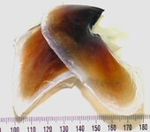
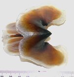
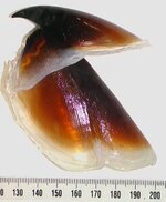
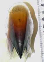
The beak, from the front, side, top, and bottom view.
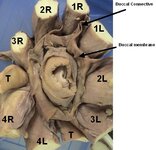
The front view showing the arrangement of tentacles and arms around the beak.
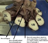
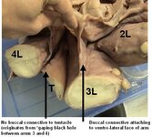
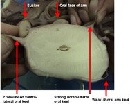
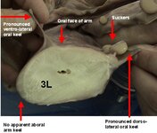
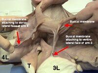
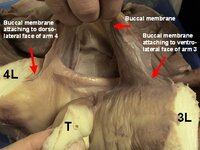
Images of the arms and tentacles illustrating their cross-sectional shapes and relative lengths. If a keel is evident on the 3rd arm, an image showing that would be great. Images showing the oral and aboral surfaces of an arm and a tentacle stretched out would also help. These can also be part of a composite panoramic image detailed below.
Dorsal and ventral views of the head, mantle, and tail fins. These can also be part of a composite panoramic image detailed below.
Panoramic shots:
(These may take the place of some of the mantle and arm/tentacle images listed above)
Keeping the camera at a constant distance above the specimen, take a series of overlapping pictures down the entire length of the specimen. These images can be stitched together in a program I have called Panorama Factory to make a single long scaled image of the architeuthis without parallax distortion. Overlapping the individual images by about 20 or 33% should be sufficient. If the arms and tentacles could be arranged so those on one side of the body had the oral surface facing the camera down their length, and those on the other side had the aboral surface facing the camera, that would really maximize the information in this long composite image. Aside from being of use as a modeling and texturing reference, I'm sure that a lot of people would be interested in this image in its own right, and I’d post the finished composite image here.
How the Images Will be Used
The pictures will be serving a twofold purpose – modeling and texturing.
For the modeling aspects, the pictures will serve as references to help me get the proportions correct and to see details that might not have been included in the images I’ve seen so far.
For the texturing aspects, at the very least, I’ll be using the images as a reference to paint the coloration onto images that will be mapped onto the model. If the specimen is in fairly good condition, I may be able to use the actual photos as the texture map for the model, although the color balance may need to be adjusted if the hue changes post mortem.
Parallax, Scale, and Color Balance Considerations
I’ve seen a lot of the photographs you’ve posted and they are excellent, so my apologies in advance if this part seems obvious. These are just things that will help maximize the usability of the images at my end.
If the images can be photographed with the camera perpendicular to the archi, that would help eliminate any parallex distortions.
The lighting I’ve seen in your images has been pretty even, without hard shadows, which is good.
If a ruler can be included in each shot to the side of animal, that will help me keep the images at the same exact scale.
Including a combination color bar with a grayscale on it next to the ruler would help me adjust the color balance to tune out any hot or cold spots in the room lighting spectrum. If you don’t have a color bar handy, I’d be more than happy to pick one up and mail it to you for this.
Well, that’s all I can think of for now. Once again, thanks very much in advance for whatever you are able to do. I’ll check the local photo shops for a color/grayscale bar and mail you one if you need it.
--Carl - hoping I'm not overstepping my bounds with all of this

That’s sounds fantastic, thanks! External views will be fine.
Here’s a wishlist, followed by some info on how I’ll be using the images, then some considerations on scaling and color balance. I’m providing that info since you may think of additional images that would be useful, but that I hadn’t thought to ask for. (basic translation: I’m a little out of my depth here)
I realize that there are a lot of images requested here, and I’ll be happy with whatever you’re able to provide. I appreciate your offer, know that you’re busy, and certainly don’t want to impose on you. I’m thinking that any images you provide will be of interest to most people here, aside from helping me with my own project.
Architeuthis Image Wishlist
The beak, from the front, side, top, and bottom view.
The front view showing the arrangement of tentacles and arms around the beak.
Images of the arms and tentacles illustrating their cross-sectional shapes and relative lengths. If a keel is evident on the 3rd arm, an image showing that would be great. Images showing the oral and aboral surfaces of an arm and a tentacle stretched out would also help. These can also be part of a composite panoramic image detailed below.
Dorsal and ventral views of the head, mantle, and tail fins. These can also be part of a composite panoramic image detailed below.
Panoramic shots:
(These may take the place of some of the mantle and arm/tentacle images listed above)
Keeping the camera at a constant distance above the specimen, take a series of overlapping pictures down the entire length of the specimen. These images can be stitched together in a program I have called Panorama Factory to make a single long scaled image of the architeuthis without parallax distortion. Overlapping the individual images by about 20 or 33% should be sufficient. If the arms and tentacles could be arranged so those on one side of the body had the oral surface facing the camera down their length, and those on the other side had the aboral surface facing the camera, that would really maximize the information in this long composite image. Aside from being of use as a modeling and texturing reference, I'm sure that a lot of people would be interested in this image in its own right, and I’d post the finished composite image here.
How the Images Will be Used
The pictures will be serving a twofold purpose – modeling and texturing.
For the modeling aspects, the pictures will serve as references to help me get the proportions correct and to see details that might not have been included in the images I’ve seen so far.
For the texturing aspects, at the very least, I’ll be using the images as a reference to paint the coloration onto images that will be mapped onto the model. If the specimen is in fairly good condition, I may be able to use the actual photos as the texture map for the model, although the color balance may need to be adjusted if the hue changes post mortem.
Parallax, Scale, and Color Balance Considerations
I’ve seen a lot of the photographs you’ve posted and they are excellent, so my apologies in advance if this part seems obvious. These are just things that will help maximize the usability of the images at my end.
If the images can be photographed with the camera perpendicular to the archi, that would help eliminate any parallex distortions.
The lighting I’ve seen in your images has been pretty even, without hard shadows, which is good.
If a ruler can be included in each shot to the side of animal, that will help me keep the images at the same exact scale.
Including a combination color bar with a grayscale on it next to the ruler would help me adjust the color balance to tune out any hot or cold spots in the room lighting spectrum. If you don’t have a color bar handy, I’d be more than happy to pick one up and mail it to you for this.
Well, that’s all I can think of for now. Once again, thanks very much in advance for whatever you are able to do. I’ll check the local photo shops for a color/grayscale bar and mail you one if you need it.
--Carl - hoping I'm not overstepping my bounds with all of this